
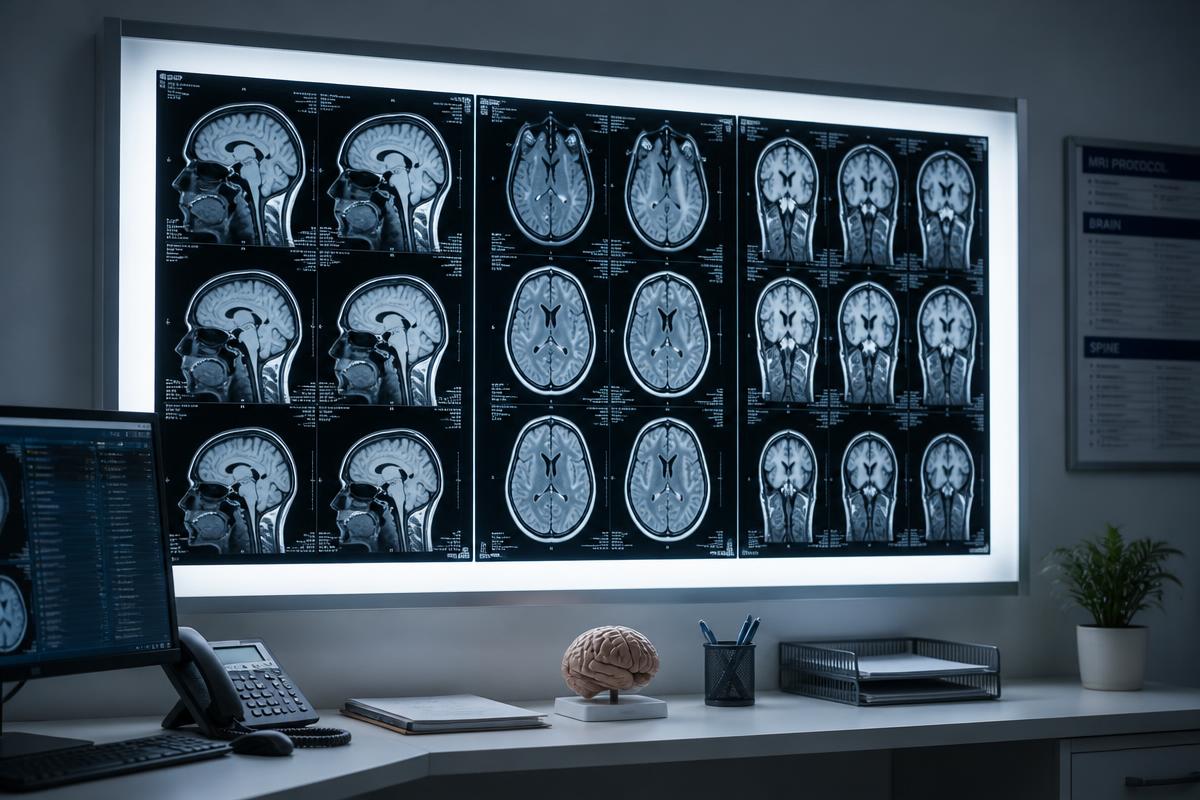
When raised intracranial pressure is suspected, imaging is usually the first test. CT and MRI reveal masses, bleeding, hydrocephalus and swelling, and they can show indirect signs of elevated pressure such as effaced sulci, compressed ventricles or an empty sella.
The limits
None of those findings is a pressure measurement. Imaging can appear reassuringly normal in a patient whose pressure is significantly elevated, and it can show chronic structural changes in a patient whose pressure is currently fine. Scans also capture a single instant, while pressure changes by the hour.
Practical implications
- A normal scan does not exclude raised intracranial pressure
- Serial scans expose patients — especially children — to repeated radiation with CT
- Imaging is expensive and often requires an emergency visit to obtain quickly
- Anatomy explains cause; pressure describes the current physiological state
Imaging and pressure monitoring answer different questions. Used together, they let clinicians see both what is structurally wrong and how the brain is coping with it right now.
← All insights